Can You Give Baby a Bath With Umbilical Cord
| Umbilical cord | |
|---|---|
 Umbilical cord of a three-infinitesimal-old baby. A medical clamp has been applied. | |
| Details | |
| Identifiers | |
| Latin | funiculus umbilicalis |
| MeSH | D014470 |
| TE | cord_by_E6.0.two.2.0.0.1 E6.0.2.2.0.0.i |
| Anatomical terminology [edit on Wikidata] | |
In placental mammals, the umbilical cord (too chosen the navel string,[1] nascence cord or funiculus umbilicalis ) is a conduit between the developing embryo or fetus and the placenta. During prenatal evolution, the umbilical string is physiologically and genetically part of the fetus and (in humans) normally contains 2 arteries (the umbilical arteries) and 1 vein (the umbilical vein), cached within Wharton's jelly. The umbilical vein supplies the fetus with oxygenated, food-rich blood from the placenta. Conversely, the fetal centre pumps low oxygen, nutrient-depleted blood through the umbilical arteries back to the placenta.
Structure and development [edit]

Cross section of the umbilical cord.

The umbilical string develops from and contains remnants of the yolk sac and allantois. Information technology forms by the fifth calendar week of evolution, replacing the yolk sac as the source of nutrients for the embryo.[ii] The string is not directly connected to the mother's circulatory system, but instead joins the placenta, which transfers materials to and from the maternal blood without allowing straight mixing. The length of the umbilical cord is approximately equal to the crown-rump length of the fetus throughout pregnancy. The umbilical cord in a full term neonate is usually almost 50 centimeters (20 in) long and most 2 centimeters (0.75 in) in bore. This diameter decreases quickly within the placenta. The fully patent umbilical artery has two main layers: an outer layer consisting of circularly arranged smooth muscle cells and an inner layer which shows rather irregularly and loosely bundled cells embedded in abundant footing substance staining metachromatic.[3] The shine muscle cells of the layer are rather poorly differentiated, contain just a few tiny myofilaments and are thereby unlikely to contribute actively to the process of post-natal closure.[iii]
Umbilical cord can be detected on ultrasound past half dozen weeks of gestation and well-visualised by 8 to 9 weeks of gestation.[4]
The umbilical cord lining is a good source of mesenchymal and epithelial stem cells. Umbilical cord mesenchymal stem cells (UC-MSC) have been used clinically to treat osteoarthritis, autoimmune diseases, and multiple other conditions. Their advantages include a ameliorate harvesting, and multiplication, and immunosuppressive properties that define their potential for utilize in transplantations. Their utilize would also overcome the ethical objections raised by the use of embryonic stem cells.[5]
The umbilical cord contains Wharton's jelly, a gelatinous substance made largely from mucopolysaccharides that protects the blood vessels inside. It contains one vein, which carries oxygenated, nutrient-rich blood to the fetus, and two arteries that carry deoxygenated, nutrient-depleted blood away.[six] Occasionally, simply two vessels (one vein and one avenue) are nowadays in the umbilical cord. This is sometimes related to fetal abnormalities, but information technology may as well occur without accompanying problems.
It is unusual for a vein to conduct oxygenated claret and for arteries to carry deoxygenated blood (the only other examples being the pulmonary veins and arteries, connecting the lungs to the eye). However, this naming convention reflects the fact that the umbilical vein carries blood towards the fetus'south centre, while the umbilical arteries carry claret abroad.
The blood flow through the umbilical cord is approximately 35 ml / min at 20 weeks, and 240 ml / min at xl weeks of gestation.[7] Adapted to the weight of the fetus, this corresponds to 115 ml / min / kg at 20 weeks and 64 ml / min / kg at twoscore weeks.[7]
For terms of location, the proximal part of an umbilical cord refers to the segment closest to the embryo or fetus in embryology and fetal medicine, and closest to the placenta in placental pathology, and opposite for the distal part, respectively.[8]
Part [edit]
Connectedness to fetal circulatory system [edit]
The umbilical cord enters the fetus via the abdomen, at the signal which (afterwards separation) will go the belly button (or umbilicus). Inside the fetus, the umbilical vein continues towards the transverse crevice of the liver, where information technology splits into two. One of these branches joins with the hepatic portal vein (connecting to its left branch), which carries blood into the liver. The 2d branch (known every bit the ductus venosus) bypasses the liver and flows into the inferior vena cava, which carries claret towards the heart. The two umbilical arteries branch from the internal iliac arteries and pass on either side of the urinary bladder into the umbilical cord, completing the circuit back to the placenta.[nine]
Changes later on birth [edit]
In absence of external interventions, the umbilical cord occludes physiologically presently later birth, explained both past a swelling and plummet of Wharton's jelly in response to a reduction in temperature and by vasoconstriction of the claret vessels by polish muscle contraction. In effect, a natural clench is created, halting the flow of blood. In air at 18 °C, this physiological clamping will accept three minutes or less.[10] In water birth, where the h2o temperature is close to body temperature, normal pulsation can exist 5 minutes and longer.
Closure of the umbilical artery by vasoconstriction consists of multiple constrictions which increase in number and degree with time. There are segments of dilatations with trapped uncoagulated blood between the constrictions before complete apoplexy.[11] Both the partial constrictions and the ultimate closure are mainly produced by musculus cells of the outer circular layer.[3] In contrast, the inner layer seems to serve mainly every bit a plastic tissue which can easily be shifted in an axial direction and and so folded into the narrowing lumen to complete the closure.[3] The vasoconstrictive apoplexy appears to be mainly mediated past serotonin[12] [13] and thromboxane Aii.[12] The artery in cords of preterm infants contracts more to angiotensin Ii and arachidonic acid and is more sensitive to oxytocin than in term ones.[13] In contrast to the contribution of Wharton's jelly, cooling causes only temporary vasoconstriction.[13]
Within the child, the umbilical vein and ductus venosus close upwards, and degenerate into gristly remnants known as the round ligament of the liver and the ligamentum venosum respectively. Part of each umbilical artery closes up (degenerating into what are known equally the medial umbilical ligaments), while the remaining sections are retained every bit part of the circulatory organization.
Clinical significance [edit]
Issues and abnormalities [edit]

A knotted cord on a newborn baby
A number of abnormalities can touch on the umbilical cord, which tin crusade problems that affect both mother and child:[14]
- Umbilical cord compression can effect from, for example, entanglement of the cord,[15] a knot in the cord,[fifteen] or a nuchal cord,[15] (which is the wrapping of the umbilical string around the fetal neck)[16] but these conditions exercise not e'er cause obstacle of fetal circulation.
- Velamentous cord insertion
- Unmarried umbilical artery
- Umbilical cord prolapse
- Vasa praevia
Clamping and cutting [edit]
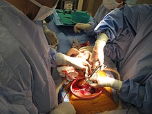
The umbilical cord is about to be cutting with pair of scissors via cesarean section

A day-old baby with its cord stump notwithstanding attached.

A 7 cm (2.75 in) long detached umbilical cord.
The cord can exist clamped at different times; nevertheless, delaying the clamping of the umbilical cord until at least one minute after birth improves outcomes equally long every bit there is the ability to care for the small hazard of jaundice if it occurs.[17] Clamping is followed by cutting of the cord, which is painless due to the absence of nerves. The string is extremely tough, similar thick sinew, and so cutting it requires a suitably sharp instrument. While umbilical severance may be delayed until afterwards the cord has stopped pulsing (one–3 minutes after nascence), there is unremarkably no pregnant loss of either venous or arterial blood while cutting the cord. Electric current evidence neither supports, nor refutes, delayed cutting of the cord, according to the American Congress of Obstetricians and Gynecologists (ACOG) guidelines.
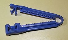
In that location are umbilical string clamps which incorporate a pocketknife. These clamps are safer and faster, allowing one to first utilize the cord clench so cut the umbilical cord. Subsequently the cord is clamped and cut, the newborn wears a plastic clip on the umbilicus area until the compressed region of the cord has dried and sealed sufficiently.
The length of umbilical left fastened to the newborn varies by practise; in most infirmary settings the length of cord left attached later clamping and cutting is minimal. In the United states, nonetheless, where the nativity occurred outside of the hospital and an emergency medical technician (EMT) clamps and cuts the cord, a longer segment up to 18 cm (vii in) in length[18] [nineteen] is left attached to the newborn.
The remaining umbilical stub remains for up to x days every bit it dries and then falls off.
Early versus delayed clamping [edit]
A Cochrane review in 2013 came to the conclusion that delayed cord clamping (betwixt one and three minutes later birth) is "likely to be beneficial every bit long as access to treatment for jaundice requiring phototherapy is available".[20] In this review delayed clamping, as contrasted to early, resulted in no difference in risk of severe maternal postpartum hemorrhage or neonatal mortality, and a depression Apgar score. On the other hand, delayed clamping resulted in an increased birth weight of on average most 100 1000, and an increased hemoglobin concentration of on average i.5 1000/dL with half the risk of beingness iron deficient at iii and six months, but an increased chance of jaundice requiring phototherapy.[20]
In 2012, the American Higher of Obstetricians and Gynecologists officially endorsed delaying clamping of the umbilical cord for 30–60 seconds with the newborn held beneath the level of the placenta in all cases of preterm commitment based largely on evidence that it reduces the risk of intraventricular hemorrhage in these children by fifty%.[21] [ obsolete source ] In the same committee statement, ACOG likewise recognize several other probable benefits for preterm infants, including "improved transitional circulation, better institution of cerise blood cell volume, and decreased need for blood transfusion". In January 2017, a revised Committee Opinion extended the recommendation to term infants, citing data that term infants benefit from increased hemoglobin levels in the newborn flow and improved iron stores in the beginning months of life, which may result in improved developmental outcomes. ACOG recognized a small increase in the incidence of jaundice in term infants with delayed cord clamping, and recommended policies exist in place to monitor for and treat neonatal jaundice. ACOG likewise noted that delayed cord clamping is not associated with increased risk of postpartum hemorrhage.[22]
Several studies have shown benefits of delayed string clamping: A meta-analysis[23] showed that delaying clamping of the umbilical cord in full-term neonates for a minimum of ii minutes post-obit birth is benign to the newborn in giving improved hematocrit, iron status as measured past ferritin concentration and stored atomic number 26, too as a reduction in the risk of anemia (relative gamble, 0.53; 95% CI, 0.xl–0.70).[23] A decrease was also found in a study from 2008.[24] Although in that location is higher hemoglobin level at 2 months, this result did not persist beyond 6 months of historic period.[25] Not clamping the string for three minutes following the birth of a baby improved outcomes at iv years of age.[26] A delay of three minutes or more in umbilical cord clamping after birth reduce the prevalence of anemia in infants.[27]
Negative effects of delayed cord clamping include an increased run a risk of polycythemia. Still, this condition appeared to be beneficial in studies.[23] Infants whose cord clamping occurred later on than sixty seconds later on birth had a higher charge per unit of neonatal jaundice requiring phototherapy.[25]
Delayed clamping is non recommended as a response to cases where the newborn is not animate well and needs resuscitation. Rather, the recommendation is instead to immediately clamp and cut the cord and perform cardiopulmonary resuscitation.[28] The umbilical string pulsating is not a guarantee that the babe is receiving enough oxygen.[29]
Umbilical nonseverance [edit]
Some parents choose to omit string severance entirely, a practice called "lotus birth" or umbilical nonseverance. The unabridged intact umbilical string is allowed to dry and separates on its own (typically on the 3rd day after birth), falling off and leaving a healed omphalos.[xxx]
Umbilical cord catheterization [edit]
As the umbilical vein is directly continued to the cardinal circulation, it can be used as a road for placement of a venous catheter for infusion and medication. The umbilical vein catheter is a reliable culling to percutaneous peripheral or primal venous catheters or intraosseous canulas and may exist employed in resuscitation or intensive intendance of the newborn.
Blood sampling [edit]
From 24 to 34 weeks of gestation, when the fetus is typically viable, blood can be taken from the string in society to test for abnormalities (particularly for hereditary conditions). This diagnostic genetic exam procedure is known as percutaneous umbilical cord blood sampling.[31]
Storage of cord blood [edit]
The blood inside the umbilical cord, known equally cord blood, is a rich and readily available source of primitive, undifferentiated stem cells (of type CD34-positive and CD38-negative). These cord claret cells can be used for os marrow transplant.
Some parents choose to accept this blood diverted from the baby's umbilical claret transfer through early cord clamping and cutting, to freeze for long-term storage at a cord claret bank should the child always require the string blood stem cells (for example, to replace os marrow destroyed when treating leukemia). This practice is controversial, with critics asserting that early on cord blood withdrawal at the fourth dimension of birth actually increases the likelihood of childhood affliction, due to the high book of blood taken (an boilerplate of 108ml) in relation to the baby's total supply (typically 300ml).[24] The Imperial College of Obstetricians and Gynaecologists stated in 2006 that "at that place is nonetheless insufficient evidence to recommend directed commercial cord blood collection and stalk-cell storage in depression-risk families".[32]
The American Academy of Pediatrics has stated that cord blood banking for self-use should be discouraged (equally most weather requiring the use of stalk cells will already exist in the cord blood), while cyberbanking for general employ should exist encouraged.[33] In the future, cord blood-derived embryonic-like stem cells (CBEs) may be banked and matched with other patients, much like claret and transplanted tissues. The employ of CBEs could potentially eliminate the upstanding difficulties associated with embryonic stalk cells (ESCs).[34]
While the American Academy of Pediatrics discourages individual banking except in the case of existing medical need, it likewise says that data near the potential benefits and limitations of cord blood banking and transplantation should be provided so that parents tin can make an informed decision.
In the United States, cord blood instruction has been supported by legislators at the federal and land levels. In 2005, the National University of Sciences published an Institute of Medicine (IoM) written report which recommended that expectant parents exist given a counterbalanced perspective on their options for cord claret banking. In response to their constituents, state legislators across the state are introducing legislation intended to help inform physicians and expectant parents on the options for altruistic, discarding or banking lifesaving newborn stalk cells. Currently 17 states, representing two-thirds of U.S. births, have enacted legislation recommended by the IoM guidelines.
The use of cord blood stem cells in treating conditions such equally brain injury[35] and Type 1 Diabetes[36] is already being studied in humans, and earlier stage research is being conducted for treatments of stroke,[37] [38] and hearing loss.[39]
String blood stored with private banks is typically reserved for apply of the donor child only. In dissimilarity, string blood stored in public banks is accessible to anyone with a closely matching tissue type and demonstrated need.[40] The use of cord blood from public banks is increasing. Currently it is used in identify of a bone marrow transplant in the treatment of blood disorders such every bit leukemia, with donations released for transplant through one registry, Netcord.org,[41] passing i,000,000 every bit of January 2013. Cord blood is used when the patient cannot find a matching bone marrow donor; this "extension" of the donor pool has driven the expansion of public banks.
The umbilical cord in other animals [edit]
The umbilical string in some mammals, including cattle and sheep, contains ii distinct umbilical veins. There is only one umbilical vein in the human umbilical string.[42]
In some animals, the mother will gnaw through the string, thus separating the placenta from the offspring. The cord along with the placenta is often eaten by the mother, to provide nourishment and to dispose of tissues that would otherwise attract scavengers or predators.[ citation needed ] In chimpanzees, the mother leaves the cord in place and nurses her immature with the cord and placenta attached until the cord dries out and separates naturally, within a day of birth, at which time the cord is discarded. (This was start documented by zoologists in the wild in 1974.[43])
Some species of shark—hammerheads, requiems and smooth-hounds—are viviparous and take an umbilical cord attached to their placenta.[44]
Animals that lay eggs seem to have a false umbilical-string that attaches the embryo and yolk together in much the same way.[ citation needed ]
Other uses for the term "umbilical cord" [edit]
The term "umbilical cord" or just "umbilical" has likewise come up to be used for other cords with similar functions, such as the hose connecting surface-supplied divers to their surface supply of air and/or heating, or space-suited astronauts to their spacecraft. Engineers sometimes use the term to describe a circuitous or critical cablevision connecting a component, particularly when composed of bundles of conductors of dissimilar colors, thickness and types, terminating in a single multi-contact disconnect.
Cancer-causing toxicants in homo umbilical cords [edit]
In multiple American and international studies, cancer-causing chemicals have been found in the blood of umbilical cords. These originate from certain plastics, figurer circuit boards, fumes and synthetic fragrances amid others. Over 300 chemic toxicants take been constitute, including bisphenol A (BPA), tetrabromobisphenol A (TBBPA), Teflon-related perfluorooctanoic acid, galaxolide and constructed musks amid others.[45] The studies in America showed college levels in African Americans, Hispanic Americans and Asian Americans due, it is thought, to living in areas of higher pollution.[46]
Additional images [edit]
-

Exclusive program of the gravid uterus in the third and fourth month.
-

Fetus in utero, between 5th and sixth months.
-
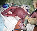
A newborn at 45 seconds, with umbilical string clamped.
-

Umbilic. Deep dissection. Serial cantankerous section.
-

dried umbilical cord stump.
See also [edit]
- Navel
- Umbilical line
References [edit]
- ^ "Umbilical – Search Online Etymology Dictionary". www.etymonline.com. Archived from the original on 4 March 2016. Retrieved 27 March 2018.
- ^ "The Umbilical Cord". yale.edu. Archived from the original on 28 March 2013. Retrieved 27 March 2018.
- ^ a b c d Meyer WW, Rumpelt HJ, Yao Air conditioning, Lind J (July 1978). "Construction and closure mechanism of the human umbilical artery". Eur. J. Pediatr. 128 (iv): 247–59. doi:x.1007/BF00445610. PMID 668732. S2CID 37516644.
- ^ Moshiri, Mariam; Zaidi, Sadaf F.; Robinson, Tracy J.; Bhargava, Puneet; Siebert, Joseph R.; Dubinsky, Theodore J.; Katz, Douglas S. (January 2014). "Comprehensive Imaging Review of Abnormalities of the Umbilical Cord". RadioGraphics. 34 (1): 179–196. doi:x.1148/rg.341125127. ISSN 0271-5333.
- ^ Saleh, R; Reza, HM (10 Oct 2017). "Short review on human umbilical cord lining epithelial cells and their potential clinical applications". Stem Cell Research & Therapy. 8 (1): 222. doi:10.1186/s13287-017-0679-y. PMC5634865. PMID 29017529.
- ^ "Fetal Circulation". www.heart.org. Archived from the original on 22 December 2017. Retrieved 27 March 2018.
- ^ a b Kiserud, T.; Acharya, K. (2004). "The fetal circulation". Prenatal Diagnosis. 24 (thirteen): 1049–1059. doi:10.1002/pd.1062. PMID 15614842. S2CID 25040285.
- ^ Mikael Häggström, Dr. (2020-08-27). Patholines https://patholines.org/Placenta.
- ^ "Peculiarities in the Vascular System in the Fetus – Gray'south Anatomy of the Homo Body – Yahoo! Educational activity". Archived from the original on January 23, 2012.
- ^ Cohain, J. S. (2010). "A Proposed Protocol for Third Phase Direction – Judy's three,4,5,x minute method". Nascence. 37 (1): 84–85. doi:10.1111/j.1523-536x.2009.00385_2.x. PMID 20402731.
- ^ Yao Ac, Lind J, Lu T (1977). "Closure of the human umbilical artery: a physiological sit-in of Burton'due south theory". Eur. J. Obstet. Gynecol. Reprod. Biol. 7 (6): 365–eight. doi:x.1016/0028-2243(77)90064-viii. PMID 264063.
- ^ a b Quan A, Leung SW, Lao TT, Man RY (Dec 2003). "v-hydroxytryptamine and thromboxane A2 as physiologic mediators of human umbilical avenue closure". J. Soc. Gynecol. Investig. 10 (8): 490–5. doi:x.1016/S1071-5576(03)00149-7. PMID 14662162.
- ^ a b c White RP (January 1989). "Pharmacodynamic study of maturation and closure of homo umbilical arteries". Am. J. Obstet. Gynecol. 160 (1): 229–37. doi:10.1016/0002-9378(89)90127-0. PMID 2912087.
- ^ "Umbilical Cord Complications: eMedicine Obstetrics and Gynecology". Archived from the original on 2010-01-29. Retrieved 2010-01-24 .
- ^ a b c P02.5 Fetus and newborn afflicted by other compression of umbilical string Archived 2014-11-02 at the Wayback Machine in ICD-10, the International Statistical Classification of Diseases
- ^ "Fetus or Newborn Problems: Labor and Delivery Complications: Merck Manual Home Edition". Archived from the original on 2010-03-28. Retrieved 2010-03-27 .
- ^ McDonald, SJ; Middleton, P; Dowswell, T; Morris, PS (Jul 11, 2013). "Effect of timing of umbilical cord clamping of term infants on maternal and neonatal outcomes". The Cochrane Database of Systematic Reviews. 7 (seven): CD004074. doi:10.1002/14651858.CD004074.pub3. PMC6544813. PMID 23843134.
- ^ Stiegler, Paul M., Thousand.D. (May 2007). "EMT-BASIC \ EMT BASIC INTERMEDIATE TECHNICIAN PROTOCOLS" (PDF). Dane Canton Emergency Medical Services. Archived (PDF) from the original on Apr half-dozen, 2012. Retrieved Dec 17, 2011.
{{cite web}}: CS1 maint: multiple names: authors listing (link) - ^ Meersman, Jack. "EMT-B: Obstetrics, Infants and Children". Wild Iris Medical Education. Archived from the original on January 20, 2012. Retrieved December 17, 2011.
- ^ a b McDonald, S. J.; Middleton, P.; Dowswell, T.; Morris, P. S. (2013). McDonald, Susan J (ed.). "Effect of timing of umbilical cord clamping of term infants on maternal and neonatal outcomes". The Cochrane Database of Systematic Reviews. 7 (7): CD004074. doi:10.1002/14651858.CD004074.pub3. PMC6544813. PMID 23843134.
- ^ Committee on Obstetric Exercise, American Higher of Obstetricians and, Gynecologists (December 2012). "Commission Opinion No.543: Timing of umbilical cord clamping subsequently nascence". Obstetrics and Gynecology. 120 (6): 1522–6. doi:10.1097/01.aog.0000423817.47165.48. PMID 23168790.
- ^ Committee on Obstetric Practise (2017-01-01). "Committee Opinion No. 684". Obstetrics & Gynecology. 129 (1): e5–e10. doi:10.1097/aog.0000000000001860. ISSN 1873-233X. PMID 28002310.
- ^ a b c Hutton EK, Hassan ES (March 2007). "Tardily vs early clamping of the umbilical cord in full-term neonates: systematic review and meta-analysis of controlled trials". JAMA. 297 (11): 1241–52. doi:x.1001/jama.297.11.1241. PMID 17374818.
- ^ a b Test of the Newborn & Neonatal Health: A Multidimensional Arroyo, p. 116-117
- ^ a b "Result of timing of umbilical cord clamping of term infants on maternal and neonatal outcomes." Cochrane Database Syst Rev. 2008; (2):CD004074
- ^ Andersson, O; Lindquist, B; Lindgren, M; Stjernqvist, K; Domellöf, M; Hellström-Westas, L (i July 2015). "Effect of Delayed Cord Clamping on Neurodevelopment at 4 Years of Age: A Randomized Clinical Trial". JAMA Pediatrics. 169 (seven): 631–8. doi:x.1001/jamapediatrics.2015.0358. PMID 26010418.
- ^ "Can delayed umbilical cord clamping reduce baby anemia at age 8, 12 months?". medicalxpress.com. Archived from the original on 25 February 2018. Retrieved 27 March 2018.
- ^ Military Obstetrics & Gynecology – Delivery of the Babe Archived 2010-01-23 at the Wayback Automobile The Brookside Associates Medical Education Division. Retrieved Jan x, 2009
- ^ Waterbirth International – Waterbirth FAQ Retrieved Jan 10, 2009
- ^ Crowther, S (2006). "Lotus birth: leaving the string alone". The Practising Midwife. 9 (6): 12–14. PMID 16830839.
- ^ "Man Reproduction, Lectures: Clinical Genetics". Retrieved 2010-02-xi .
- ^ "Umbilical Cord Blood Cyberbanking" (PDF) (Scientific Bear on Paper viii). Royal College of Obstetricians and Gynaecologists. 2006. Retrieved 2021-07-01 .
{{cite web}}: CS1 maint: url-status (link) - ^ American Academy of Pediatrics. "Cord Blood Banking for Potential Hereafter Transplantation". Archived from the original on 2007-10-13.
- ^ "Cord blood yields 'ethical' embryonic stem cells." Archived 2008-10-10 at the Wayback Motorcar, Coghlin A. New Scientist, Baronial 18, 2005. Accessed June 25, 2007.
- ^ Cord Blood for Neonatal Hypoxic-Ischemic Encephalopathy Archived 2011-08-12 at Wikiwix, Autologous Cord Blood Cells for Hypoxic Ischemic Encephalopathy Study 1. Stage I Study of Feasibility and Condom
- ^ Haller MJ; Viener, HL; Wasserfall, C; Brusko, T; Atkinson, MA; Schatz, DA; et al. (2008). "Autologous Umbilical Cord Claret Infusion for Type ane Diabetes". Exp. Hematol. 36 (half-dozen): 710–715. doi:10.1016/j.exphem.2008.01.009. PMC2444031. PMID 18358588.
- ^ Vendrame M, et al. (2006). "String blood rescues stroke-induced changes in splenocyte phenotype and function". Exp. Neurol. 199 (one): 191–200. doi:10.1016/j.expneurol.2006.03.017. PMID 16713598. S2CID 29804539.
- ^ Vendrame M, et al. (2005). "Anti-inflammatory furnishings of human cord blood cells in a rat model of stroke". Stem Cells Dev. fourteen (five): 595–604. doi:10.1089/scd.2005.14.595. PMID 16305344.
- ^ Revoltella RP, et al. (2008). "Cochlear repair by transplantation of human cord claret CD133+ cells to nod-scid mice fabricated deaf with kanamycin and racket". Cell Transplant. 17 (half dozen): 665–678. doi:10.3727/096368908786092685. PMID 18819255.
- ^ Najjar, Dana (2020-12-18). "Should Yous Bank Your Baby's Cord Blood?". The New York Times. ISSN 0362-4331. Retrieved 2021-07-01 .
- ^ "Archived copy" (PDF). Archived (PDF) from the original on 2013-xi-02. Retrieved 2013-04-13 .
{{cite web}}: CS1 maint: archived copy as title (link) - ^ Meat Hygiene. J. F. Gracey, D. South. Collins, Robert J. Huey. p. 32.
- ^ Run across In the Shadow of Man, past Jane Goodall.
- ^ "Sharks (Chondrichthyes)". FAO. Archived from the original on 2008-08-02. Retrieved 2009-09-14 .
- ^ Chen, Mei-Huei; Ha, Eun-Hee; Wen, Ting-Wen; Su, Yi-Ning; Lien, Guang-Wen; Chen, Chia-Yang; Chen, Pau-Chung; Hsieh, Wu-Shiun (iii August 2012). "Perfluorinated Compounds in Umbilical String Claret and Adverse Birth Outcomes". PLOS I. seven (eight): e42474. Bibcode:2012PLoSO...742474C. doi:10.1371/journal.pone.0042474. PMC3411780. PMID 22879996.
- ^ "Why Are Trace Chemicals Showing upwards in Umbilical Cord Blood?". Scientific American. Archived from the original on 2012-09-02. Retrieved 2012-09-01 .
- Marianne Messerli; et al. (December 2013). "Stem Cells from Umbilical String Wharton'southward Jelly from Preterm Birth Take Neuroglial Differentiation Potential". Reproductive Sciences. twenty (12): 1455–1464. doi:10.1177/1933719113488443. PMC3817666. PMID 23670950.
External links [edit]
-
 Media related to Umbilical cord at Wikimedia Commons
Source: https://en.wikipedia.org/wiki/Umbilical_cord
0 Response to "Can You Give Baby a Bath With Umbilical Cord"
Postar um comentário